Selective Photothermolysis, Explained Properly
Selective photothermolysis is the single idea every aesthetic laser and IPL on the market is built to obey. Learn it properly and a spec sheet stops being marketing. It turns into a list of claims you can check yourself.
What 1983 actually replaced
R. Rox Anderson and John A. Parrish published Selective photothermolysis: precise microsurgery by selective absorption of pulsed radiation in Science in 1983, volume 220, pages 524 to 527. Two demonstrations carried the argument. Pulses at 577nm lasting three ten-millionths of a second damaged cutaneous microvessels. Pulses at 351nm lasting twenty billionths of a second damaged melanosomes inside melanocytes. The claim that still matters most is about aiming: you don't have to aim. The target's own optical and thermal properties do the selecting.
Think about what that displaced. Argon lasers through the 1970s ran continuous or quasi-continuous, and the port-wine stain literature from that era reports frequent hypertrophic scarring and a substantial rate of pigment change. Light went in, heat spread sideways, and dose was the only lever anyone had. One paper turned a heating problem into a timing problem.
Choose a wavelength your target absorbs strongly, deliver it in a pulse matched to how fast the target sheds heat, and you damage the target while its neighbours stay intact.
Everything below is that sentence, taken apart.
Three chromophores, and the fight between them
A chromophore is whatever absorbs the photon. An absorption curve charts how greedily one molecule drinks each wavelength. Skin hands you three that matter commercially.
- Oxyhaemoglobin. Our engineering manuals put haemoglobin's absorption peaks at 418nm, 542nm and 577nm. Vascular work has lived in the blue-green and the yellow ever since, and that's why Anderson and Parrish reached for 577nm.
- Melanin. No tidy peak at all. Absorption simply falls as wavelength climbs, a long slide rather than a spike. Below roughly 800nm almost any wavelength preferentially heats melanin, and short light scatters hard in the epidermis. Above 800nm more of the beam gets through, so our manuals treat dermal pigment from the long side.
- Water. Barely touched between 400 and 800nm, then climbing. Published liquid-water spectra place the near-infrared bands near 970nm, 1200nm and 1450nm, with the dominant maximum in the mid-infrared: StatPearls puts water's peak near 3000nm and the 2940nm Er:YAG line closest to it. One correction, since we've seen it repeated off our own chart: Chapter 4 of our internal training text labels 980nm, 1060nm and 1480nm as water bands. Read 980 and 1480 as device wavelengths chosen to work with water, not as spectral maxima, and drop 1060 entirely. Water absorption around 1064nm is low. That trough is why Nd:YAG travels.
Here's the part sales decks skip. Your safety margin is not how strongly the target absorbs. It's the ratio between what the target absorbs and what the epidermis above it absorbs at the same wavelength. 532nm sits near a haemoglobin peak and is drunk greedily by melanin too, so it clears freckles beautifully on pale skin and burns dark skin. 1064nm is absorbed weakly by both, so it reaches dermal targets with the epidermis relatively intact. That contrast is why our QE-01 Q-switched Nd:YAG carries both 1064nm and 532nm. Between roughly 600 and 1200nm sits what our manuals call the optical window: low scattering, little pigment absorption.
Thermal relaxation time, worked through
Thermal relaxation time is how long a heated target takes to shed about 63% of its absorbed heat. Clinical texts invert it: the time for temperature to fall to 37% of peak. Same number.
Now the relation that matters. Thermal relaxation time rises with the square of the target's size. Double the diameter, quadruple the cooling time. A melanosome and a follicle aren't two settings on one dial. They're orders of magnitude apart, and so are their pulses.
| Target | Scale | Cooling time | Pulse regime that follows |
|---|---|---|---|
| Tattoo ink particle | Sub-micron pigment grain | Under 1 nanosecond in the pigmented-lesion literature | Q-switched nanoseconds. Our QE-01 runs 6ns |
| Melanosome | Organelle, around a micron | About 50 nanoseconds in one review; other texts argue closer to a microsecond | Nanoseconds. Distrust any single quoted value |
| Epidermal basal layer | Roughly 20 micrometres thick | Estimated at 1.6 to 2.8 milliseconds | Not a target. The clock your cooling must beat |
| Cutaneous microvessel | Capillary calibre | Anderson and Parrish used 0.3 microsecond pulses at 577nm | Sub-microsecond to milliseconds, by vessel size |
| Pigmented hair follicle | A structure, not an organelle | Milliseconds | Our DL-07 808nm diode platform spans 5 to 200ms |
The hair row deserves a minute. At a fixed fluence, our training material has a melanin-bearing follicle coagulating at around 100ms and the epidermis injured at around 99ms. One millisecond of margin. Epidermal protection isn't a comfort feature on a hair removal machine. It's what stands between the physics and a burn.
One refinement matters. Altshuler and colleagues extended the theory in 2001 with a thermal damage time, arguing that for unevenly pigmented targets the right pulse runs significantly longer than the target's own thermal relaxation time. Picture it. In a follicle the melanin sits in the shaft and matrix while the target cells sit elsewhere, so heat needs time to travel from absorber to victim.
Fluence, spot and cooling decide as much as wavelength
Fluence is energy per unit area, in joules per square centimetre: power multiplied by time, divided by spot area. Under the damage threshold you achieve nothing. Over the epidermal threshold you injure the layer you meant to protect. Wavelength picks the target. Fluence decides whether anything happens to it.
Spot size is the parameter buyers underrate most. At a fixed wavelength, a larger spot penetrates deeper and spreads energy more evenly, because proportionally fewer photons scatter out sideways on the way down. So 8 J/cm² at 2mm and 8 J/cm² at 6mm are not the same treatment.
The skin takes its cut first. Around 4 to 7% of light reflects straight off the surface at normal incidence, and dry skin reflects more. Hence coupling gel, and hence the cost of an angled shot.
Cooling buys headroom to push fluence at a deep target. Our manuals are blunt about the limits. If the target is epidermal pigment under a Q-switched pulse, avoid surface cooling at the instant of the pulse. For a papillary-dermis vessel, use vigorous pre- and post-treatment cooling instead. Guidance for darker skin points the same way: longer wavelengths, lower fluences, longer pulse durations.
Where the model breaks down
Selective photothermolysis is a model. Models have edges.
- Bulk heating. The theory treats one pulse in isolation. Stack pulses fast enough and tissue never returns to baseline. Our manuals say it plainly: more pulses means more pulse width, and more thermal coagulation. Repetition rate and pulse count are thermal parameters.
- Photoacoustic, not photothermal. At nanosecond scale with enormous peak power, small dense targets don't gently warm. They fragment. Reviews attribute tattoo ink clearance to photomechanical, or photoacoustic, fragmentation, and picosecond systems lean on it almost entirely. Temperature stops being the useful variable.
- The target changes while you treat it. Heated haemoglobin oxidises to methaemoglobin, which our archive notes absorbs 1064nm about 13 times more strongly than deoxyhaemoglobin and three times more than oxyhaemoglobin. Your absorption coefficient moves mid-pulse-train.
- Melanin that isn't a thin film. In higher phototypes, and across much of Asian skin, pigment sits in ways no wavelength fully sidesteps. One comparison found nanosecond treatment produced post-inflammatory hyperpigmentation in 22% of cases against 6% for a long-pulsed approach. PIH is driven by basal melanocyte activity provoked by inflammation, so a treatment that inflames can manufacture the pigment it was hired to remove. Risk runs higher across Fitzpatrick IV to VI, which is why our note on reading Fitzpatrick skin types exists.
- Physics sets a ceiling, not a promise. Even the textbook application falls short: reviews of pulsed dye laser treatment of port-wine stains report suboptimal response in 20% to 46% of patients. Diagnosis belongs to a qualified clinician before any device is switched on.
How to interrogate a spec sheet
In a sales conversation the principle becomes four questions.
- Which wavelength, and which chromophore does it serve? If the answer is a list of indications rather than an absorber, the vendor is reading you a brochure.
- What is the true pulse width, and does it move when energy moves? On an actively Q-switched laser the pulse width stays constant and changing energy changes peak power, nothing else. Anyone selling tattoo removal with a pulse quoted in microseconds is selling something else. Our QE-01 states 6ns.
- What fluence range, at what spot size? A fluence figure without its spot is unfalsifiable. Ask for maximum energy at the largest spot. The DL-07 quotes 2 to 80 J/cm² across a 13 x 15mm tip, so the two numbers travel together.
- What cooling, and does it hold through a full clinic day? A tip that warms up by the fourth client has rewritten your safety margin.
One last number for your pocket. A 200mJ Nd:YAG running free generates roughly a 400 microsecond pulse. Q-switch the same 200mJ down to 10ns and peak power rises about 40,000 times. Identical energy, different machine.
Those four questions travel well. Put them on one card and make every vendor answer in units, not adjectives.
Want ours answered? Ask. Pmise engineering will send the measured pulse width and the fluence-at-spot documentation for the QE-01 and the DL-07 on request through our contact page. Two things distributors raise next: operator training runs remotely and covers parameters, screening and test-spot discipline, and wear parts including cooling tips are supported by factory spares under a one-year warranty, with lifetime maintenance after it.
Evidence and further reading
Educational material for equipment buyers and operators. It is not medical advice, an operating protocol or a promise of clinical outcome.
- Anderson RR, Parrish JA. Selective photothermolysis: precise microsurgery by selective absorption of pulsed radiation. Science 1983;220(4596):524-527
- Altshuler GB, Anderson RR, Manstein D, Zenzie HH, Smirnov MZ. Extended theory of selective photothermolysis. Lasers Surg Med. 2001;29(5):416-432
- Carr JA et al. Absorption by water increases fluorescence image contrast of biological tissue in the shortwave infrared. PNAS 2018;115(37):9080-9085 (water absorption peaks near 970, 1200 and 1450nm)
- StatPearls: Laser Erbium-YAG Resurfacing, 2940nm and the water absorption peak (NCBI Bookshelf)
- Theoretical review of the treatment of pigmented lesions in Asian skin (thermal relaxation times, photomechanical fragmentation, PIH rates)
- Laser treatment of port-wine stains: argon-era scarring and pigmentation risk, and modern response rates
- StatPearls: Laser Fitzpatrick Skin Type Recommendations (NCBI Bookshelf)
- StatPearls: Intense Pulsed Light (IPL) Therapy, thermal relaxation time and chromophores (NCBI Bookshelf)
Put it to work
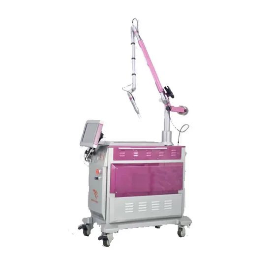 The machine that fitsPmise QE-01
EO Q-Switched Nd:YAG, 1064/532nm Next noteIPL vs Laser: How to Choose for Your Clinic
Filtered broadband light against a single coherent wavelength. Coverage, versatility, cost per treatment and the filter system that keeps UV off the skin. CatalogAll platforms
Twelve machines, one factory standard, full specifications.
Ready to spec your next machine?
Tell us the indication, your market and the certifications you need. Our export team replies with a configuration and quotation within 12 to 24 hours.